Ultrasound Paracentesis Trainer
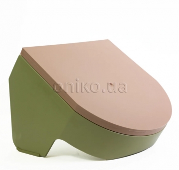
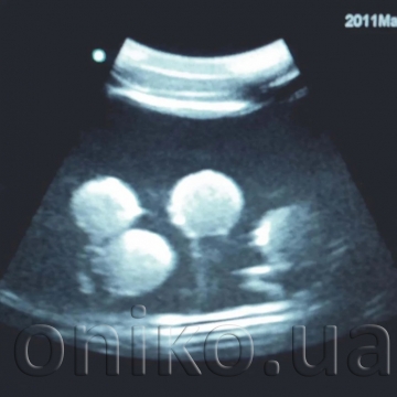
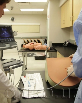
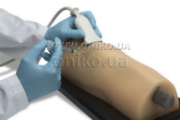
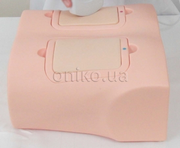
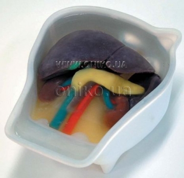
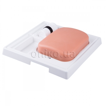
Ultrasound Paracentesis Trainer ultrasound compatible trainer allows the user to perform diagnostic and/or therapeutic paracentesis. The anatomically correct trainer is positioned to simulate a patient sitting up at a 45-degree angle with a distended abdomen and includes the pubic symphysis, iliac crest, and umbilicus. The inferior epigastric vessels can be visualized under ultrasound.
Features:
- Ultrasound compatible with replaceable tissue
- Internal anatomy includes superficial epigastric vessels, liver and spleen, rectus abdominal muscles, and intestines
- Allows up to one liter of intraperitoneal fluid removal
- Replaceable tissue is durable and allows for repeated use
- Palpable anatomy and realistic needle response
Skills:
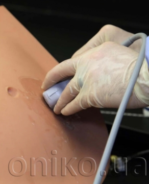
- Use an ultrasound probe to minimize risks and learn to:
- Locate the pocket of ascites
- Gauge the distance from skin to fluid, and avoid bowel
- Look for blood vessels beneath the skin using a vascular probe
- Drain the fluid from the peritoneal cavity
- Use the traditional lateral gutter technique or midline linea alba technique
- Palpate anatomic landmarks significant to the procedure

