Vascular Access and Regional Anesthesia Ultrasound Training Model
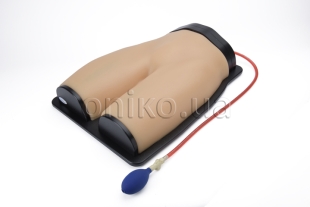
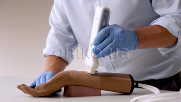
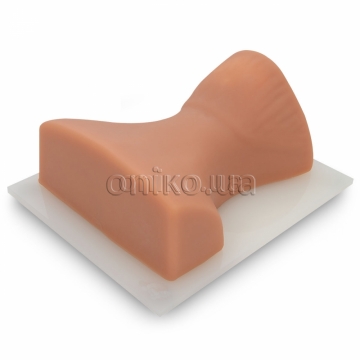
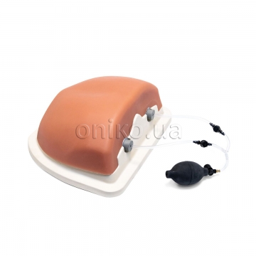
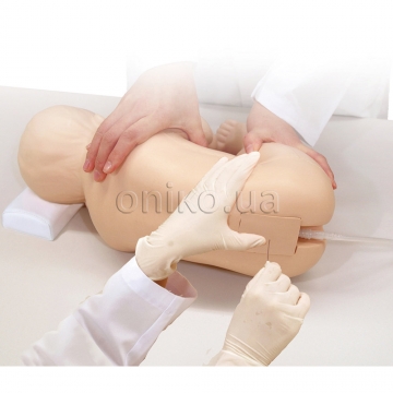
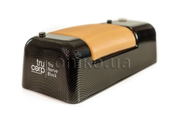
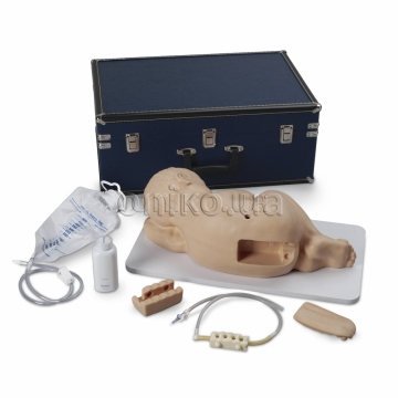
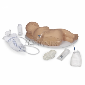
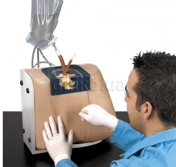
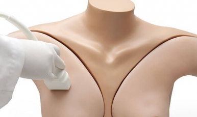
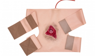
Generation II Femoral lower torso ultrasound guided regional anesthesia and central venous access training model is an excellent tool to train users to develop and practice the skills necessary to gain proficiency in using ultrasound for regional anesthesia procedures of the femoral nerve as well as guide catheter insertions in the femoral artery and vein. All of our simulation materials match the acoustic characteristics of real human tissue so when you use your ultrasound system on our training models, you experience the same quality you expect from imaging patients in a clinical environment. Ultra-durable, self-healing fabric that feels like human skin will allow you to perform local anesthetic blocks and central venous access procedures repeatedly.
The model offers an anatomically correct femoral nerve as well as fascia of the iliacus nerve. Vascular anatomy includes: aorta, inferior vena cava (IVC), common iliac artery and vein, internal iliac artery and vein, femoral artery and vein, great saphenous vein. Users can use traditional anatomical landmarks such as the pubic symphysis, iliac crest, and inguinal ligament to establish blind venous access, or use ultrasound to obtain images of relevant anatomical structures.
