Intramuscular Injection Simulator
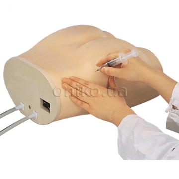
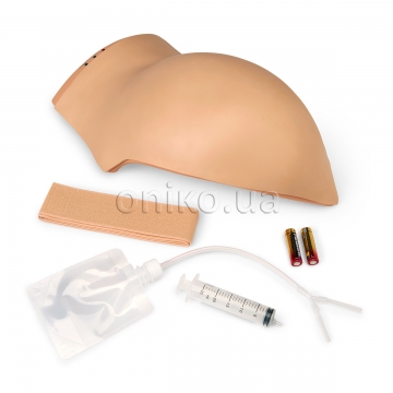
Here is the perfect volunteer for training your beginning healthcare students to administer intramuscular
injections. Always available for class demonstrations or practice by groups of any size, the Intramuscular
Injection Simulator offers both visual and tactile learning. A simulated bony structure is embedded in the
torso and represents the superior end of the femur, or greater trochanter, the posterior superior and anterior
superior iliac spines, and the sacrum. The bony structure provides palpable anatomical landmarks so
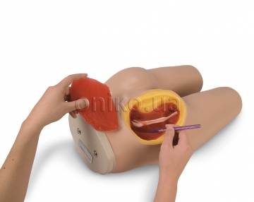
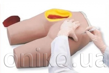
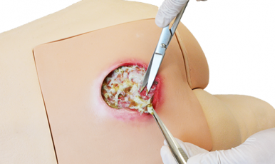
students can identify proper injection sites. A section of the upper, outer quadrant of the left gluteal area is
cut away to allow students to visualize the underlying structures. The gluteus medius and gluteus maximus
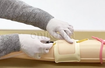
muscles, sciatic nerve, and vascular structures are clearly shown. Three types of intramuscular injections
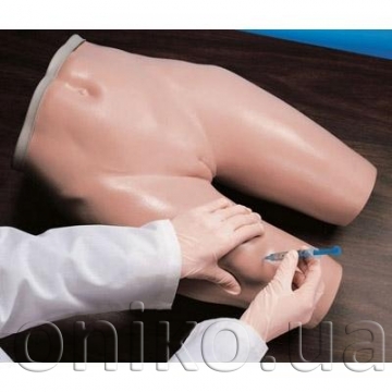
can be taught and practiced on this simulator: dorsogluteal, ventrogluteal, and vastus lateralis injections. As
with all replicas and simulators, the Intramuscular Injection Simulator is extremely realistic. Skin and
muscle textures, as well as bone shape and position, closely resemble a live patient. As a result, perforation
of the tissue with a needle duplicates the sensation of administering an actual injection.

