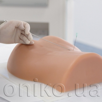
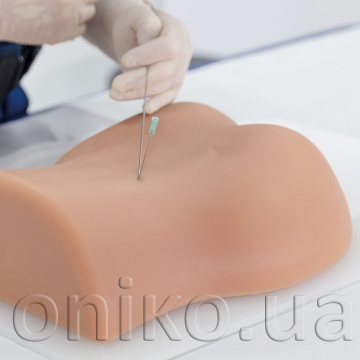
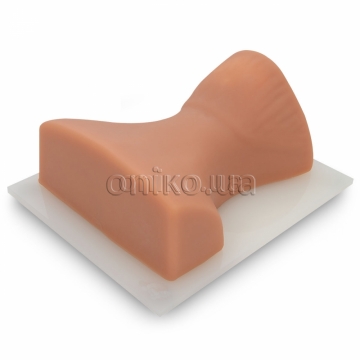
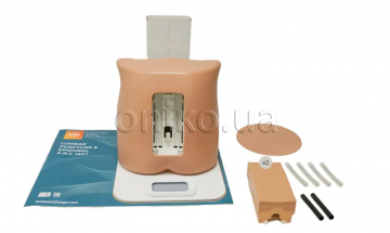
Image Guided Lumbar Spinal Injection Trainer
Improve outcomes of spine interventions with standardized image guided injection training
The Image Guided Lumbar Spinal Injection Trainer enables trainees to develop a three-dimensional understanding of the procedures for successful spine interventions. They will learn to correlate imaging with the lumbar spinal anatomy relevant to interventional pain procedures, to identify the target tissue and vulnerable structures using imaging and anatomic inspection, and to apply best practices to ensure patient safety during the interventional spine procedures.
Using the Spinal Injection Trainer is an economic alternative for imaging techniques courses on cadavers and offers the instructors a reliable, standardized patient simulation always ready to use:
• Life-like radiopacity for realistic x-Ray images
• Realistic injection haptics
• Anatomically accurate bone structure
• Visually identifiable landmarks
The following image guided spinal interventions can be trained on the simulator:
• Transforaminal Epidural Steroid Injections (TFSI)
• Intralaminar Epidural Injection
• Facet Blocks
• Medial Branch Block (MBB/RF)
• S1-block
• Sacroiliac Joint Injection (SIJ-injection)
The Lumbar Spinal Injection Trainer depicts the following bone structures:
• Sacrum, S1-S3 (with sacral hiatus canal)
• Ilium, bilateral without ischial bone and hip joint
• T12-L5
• Coccyx
The trainer is especially suitable for use in imaging techniques courses thanks to these simulation features:
• Self-Sealing material can be used repeatedly for injection training
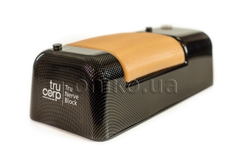
• Completely portable with a secure transport box
• Durable construction, easy to clean

